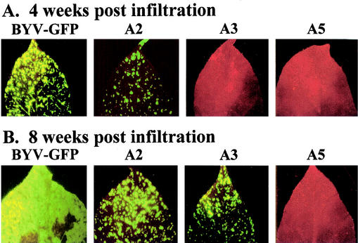
FIG. 3.
Systemic transport of the parental BYV-GFP and selected mutant variants. The noninoculated upper leaves of the N. benthamiana plants are shown. The leaves were photographed with an epifluorescence microscope at 4 (A) or 8 (B) weeks postinfiltration. The virus-infected areas are green due to GFP fluorescence, whereas the red color corresponds to noninfected, autofluorescent areas.

