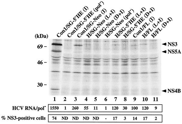
FIG. 4.
Detection of HCV proteins and RNA in Huh-7.5 cells transiently transfected with subgenomic and full-length HCV RNAs. (Top) Ninety-six hours after RNA transfection of Huh-7.5 cells, the monolayers were labeled with 35S protein-labeling mixture and lysed, and NS3, NS4B, and NS5A were analyzed by immunoprecipitation, SDS- 10% PAGE, and autoradiography. The positions of the molecular-mass standards are given on the left, and HCV-specific proteins are indicated on the right. (Middle) Total cellular RNA was extracted 96 h posttransfection, and HCV RNA levels were quantified as described in Materials and Methods. The ratio of HCV RNA to the pol− negative control is shown (HCV RNA/pol−). (Bottom) Ninety-six hours after transfection, cells were fixed with 4% paraformaldehyde, permeabilized with 0.1% saponin, stained for HCV NS3, and analyzed by FACS. The percentages of cells expressing NS3 relative to an isotype-matched irrelevant IgG are displayed. Values of <1% were considered negative (−). ND, not determined.

