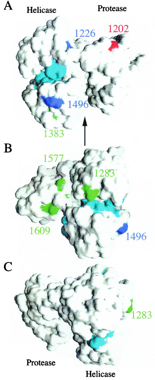
FIG. 6.
Locations of NS3 adaptive mutations. Solvent-accessible surface of the NS3/4A crystal structure (23) highlighting the locations of several adaptive mutations. Adaptive mutations described in this paper are colored blue, while published mutations from references 13 and 16 are colored red and green, respectively. The seven conservedmotifs of the RNA helicase are colored cyan. The numbering corresponds to the genotype 1 sequences. (A) The NS4A peptide and protease domain are on the right, with the helicase domain on the left. (B and C) Rotations (90 and 180°, respectively) about a vertical axis (represented by the arrow in panel A).

