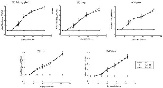
FIG. 6.
Titers of MCMV mutants in the salivary glands (A), lungs (B), spleens (C), livers (D), and kidneys (E) of the infected SCID mice. CB17 SCID mice were infected intraperitoneally with 104 PFU of each virus. At 1, 3, 7, 10, 14, and 21 days postinfection, the animals (three mice per group) were sacrificed, and the organs were collected and sonicated. The viral titers in the tissue homogenates were determined by standard plaque assays in NIH 3T3 cells. The viral titers represent the average obtained from triplicate experiments. The error bars indicate the standard deviation. Error bars that are not evident indicate that the standard deviation was less than or equal to the height of the symbols. The limit of detection was 10 PFU/ml of the tissue homogenate.

