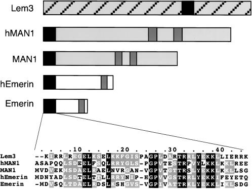
Figure 1.
Schemes of the three LEM domain proteins in C. elegans showing the positions of LEM domains (black) and transmembrane domains (dark gray). Also shown are the amino acid sequences of the LEM domains of Ce-Lem3 (Lem3), human MAN1 (hMAN), Ce-MAN1, human emerin (hEmerin), and Ce-emerin (Emerin). Numbering of the amino acids is arbitrary. Black shading indicates identical residues, gray shading indicates conserved residues, and dashes indicate gaps introduced to optimize homology.

