Figure 4.
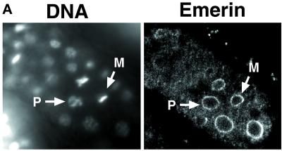
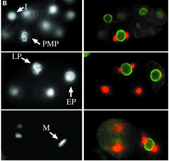
Persistence of nuclear envelope markers during mitosis in C. elegans. (A) Indirect immunofluorescence of endogenous Ce-emerin in a C. elegans embryo. DNA was stained with Hoechst 33258 (left), and the same embryo was stained for endogenous Ce-emerin with the use of mouse polyclonal serum 3272 (right; imaged by confocal microscopy). Arrows point to nuclei in prophase (P) and metaphase (M). (B) C. elegans embryos triple stained for DNA (Hoechst 33258, left) and by indirect immunofluorescence with the use of antibodies against Ce-lamin (green) and tubulin (red; right; imaged by confocal microscopy). The stages of mitosis were determined by chromosome morphology and corroborated by tubulin staining patterns: I, interphase; PMP; prometaphase; LP, late prophase; EP, early prophase; M, metaphase.


