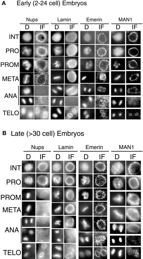
Figure 5.
Immunofluorescence localization of endogenous nuclear envelope proteins at different stages of mitosis in 2- to 24-cell C. elegans embryos (A) and >30-cell C. elegans embryos (B). Embryos were doubly stained for DNA with the use of Hoechst 33258 (D) and by indirect immunofluorescence (IF) with the use of antibodies specific for nuclear pore complexes (mAb414; Nups), Ce-lamin, Ce-emerin, or Ce-MAN1. A representative nucleus from each stage is shown: INT, interphase; PRO, prophase; PROM, prometaphase; META, metaphase; ANA, anaphase; and TELO, telophase. All Ce-emerin immunofluorescence images, plus the panel showing nucleoporin metaphase staining in early embryos and most Ce-MAN1 early embryo images, were obtained by confocal microscopy; all others were imaged by fluorescence microscopy.

