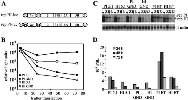
FIG. 2.
Transient replication of replicons with different IRES elements at the 5′ end. (A) Structure of the replicons rep HI-luc and rep PI-luc used for transient replication assays. 5′, HCV 5′ NTR; 3′, HCV 3′ NTR; PI, PV IRES; luc, firefly luciferase; EI, EMCV IRES. (B) Transient replication of the replicons carrying the luciferase reporter gene under translational control of the HCV IRES or the PV IRES. Huh-7 cells were transfected with 5 μg of rep PI-luc/5.1, rep PI-luc/GND, rep HI-luc/5.1, or rep HI-luc/GND, and luciferase activity was determined in cell lysates that were prepared at given time points posttransfection. Data were normalized for transfection efficiency among constructs with identical IRES elements as determined by measurement of the luciferase activity 4 h after transfection. Electroporations and luciferase assays were performed in duplicates. Note the logarithmic scale of the ordinate. (C) Analysis of transient replication of different luciferase replicons by Northern hybridization. Huh-7 cells were transfected with 5 μg of luciferase replicons 5.1 that carry two cell culture-adaptive mutations in NS3 and one in NS5A (28) or with the ET replicons carrying the same NS3 mutations and one in NS4B instead of NS5A (Table 2). Cells were harvested at different time points postelectroporation (pE) as indicated above each lane. Then, 10 μg of total RNA was analyzed by Northern hybridization with a 32P-labeled negative-sense riboprobe spanning the NS3 to NS5B region. The positions of replicon RNAs with PV IRES (rep PI) or HCV IRES (rep HI) and β-actin mRNA are given on the right. (D) Quantification of the Northern hybridization shown in panel C by phosphorimaging. Values were corrected for different RNA amounts by using the β-actin signal and normalized for the PSL obtained at 4 h after transfection.

