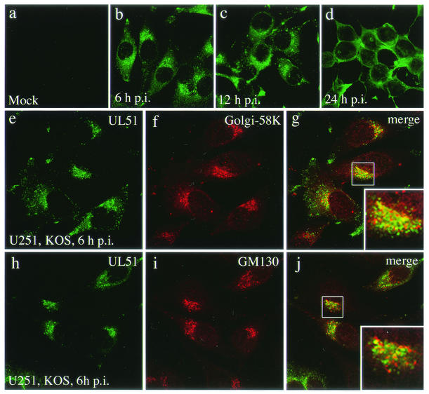
FIG. 2.
Intracellular localization of the UL51 protein in HSV-1-infected U251 cells. Mock-infected (a) and HSV-1-infected (b to j) U251 cells were fixed at 6 (b and e to j), 12 (c), and 24 (d) h p.i. as described in Materials and Methods. The samples were double stained with the anti-UL51 rabbit polyclonal antibody (a to e and h) and anti-Golgi-58K protein mouse monoclonal antibody (f) or anti-GM130 mouse monoclonal antibody (i) and then reacted with anti-rabbit IgG-conjugated FITC or anti-mouse IgG-conjugated TRITC. The merged images are shown in panels g and j. Infected cells were pretreated with normal goat serum to block nonspecific antibody reaction. The insets show high magnification of the juxtanuclear region.

