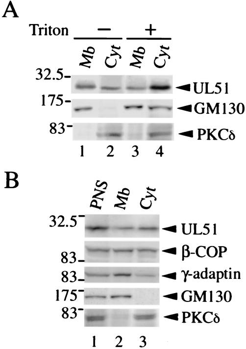
FIG. 3.
Subcellular distribution of the UL51 protein. (A) Lysates of U251 cells transiently expressing the UL51w/t protein were separated into the membranous pellet (Mb) (lanes 1 and 3) and the cytosolic supernatant (Cyt) (lanes 2 and 4) by ultracentrifugation (120,000 × g, 1 h) in the absence (lanes 1 and 2) or presence (lanes 3 and 4) of 1% Triton X-100 as described in Materials and Methods. The samples were subjected to SDS-PAGE and analyzed by Western blotting with the anti-UL51 rabbit polyclonal antibody (upper panel) and mouse monoclonal antibodies against GM130 (middle panel) or PKCδ (lower panel). (B) ST51 cells were homogenized, the resulting PNS (lane 1) was ultracentrifuged (120,000 × g, 1 h), and the membranous pellets (lane 2) and the cytosolic supernatants (lane 3) were collected. The samples were separated by SDS-PAGE and analyzed by Western blotting with the anti-UL51 rabbit polyclonal antibody and mouse monoclonal antibodies against β-COP, γ-adaptin, GM130, or PKCδ.

