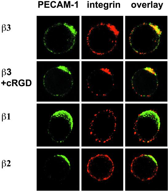
Figure 9.
Distribution of integrin αvβ3 and PECAM-1 on the surface of FTF1.26 cells. PECAM-1 cap formation and its effect on the distribution of integrin β chains on the surface of proT-cells (FTF1.26) were analyzed by confocal fluorescencse microscopy. Cell surface PECAM-1 caps (green) were induced by specific antibody cross-linking followed by detection of integrin chains β1, β2, and β3 (red). Interacting proteins colocalize together and their overlapping fluorochromes results in a yellow cap. “cRGD” indicates the addition of the cyclic RGD peptide, a ligand binding motif for integrins, during the PECAM-1 cross-linking step. Only integrin chain β3, in the absence or presence of cRGD, colocalized with PECAM-1 (yellow caps in overlay). The integrin chains β1 and β2 did not colocalize with PECAM-1 (green caps in overlay).

