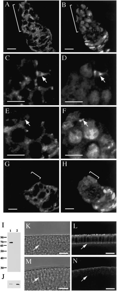
Figure 1.
Presence of Pnut in wild-type stem cells and absence of Pnut in pnut germline clones. (A–H) Immunofluorescence micrographs of wild-type (A–F) and pnut germline-clone (G and H) ovarioles, showing germaria that were double-stained with the 4C9 anti-Pnut antibody (A, C, E, and G) and with antianillin (B, D, F, and H) antibody. (A and B) Presence of Pnut in cells throughout the region of germline proliferation (indicated by brackets) in wild-type germaria. (C and D) Presence of Pnut in stem-cell cleavage furrows (arrows). (E and F) Absence of Pnut from some stem-cell ring canals (arrows). In F, note the presence of anillin in the nuclei, showing that the cells have progressed into interphase (Field and Alberts, 1995; de Cuevas and Spradling, 1998). (G and H) Absence of Pnut from the region of germline proliferation (brackets) in germline-clone germaria. The Pnut-expressing cells in G are the somatic follicle cells that surround the germline cells after germline proliferation is complete. (I–N) Absence of Pnut from germline-clone embryos. Embryos were collected from the wild-type stock and from germline-clone females that had been allowed to mate with their siblings. (I and J) Western blots of extracts of 0–3-h embryos of wild-type (lane 1) and pnut germline clones (lane 2) stained with the KEKK anti-Pnut antibody (I) or with anti-Bicaudal D antibody as a loading control (J). (K–N) Transmitted-light (K and M) and immunofluorescence (L and N) micrographs of cellularizing wild-type (K and L) and germline-clone (M and N) embryos stained with the KEKK antibody. Arrows indicate the position of the cellularization front. A weak apical background staining is seen in germline-clone embryos with this antibody. The 4C9 anti-Pnut monoclonal antibody also showed no Pnut staining at the cellularization front but had a different background pattern (our unpublished observations). Bars, 5 μm (A–H), 10 μm (K–N).

