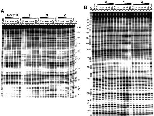
Figure 2.
Sequence selective binding. The gels show DNase I footprinting with (A) 117mer and (B) 178mer PvuII–EcoRI restriction fragments cut from the plasmids pBS and pKS, respectively. In both cases, the DNA was labeled at the EcoRI site with [α-32P]dATP in the presence of AMV reverse transcriptase. The products of nuclease digestion were resolved on an 8% polyacrylamide gel containing 7 M urea. Control tracks (Cont) contained no drug. The concentration (µM) of the drug is shown at the top of the appropriate gel lanes. Tracks labeled ‘G’ represent dimethylsulfate-piperidine markers specific for guanines. Numbers on the side of the gels refer to the standard numbering scheme for the nucleotide sequence of the DNA fragment.

