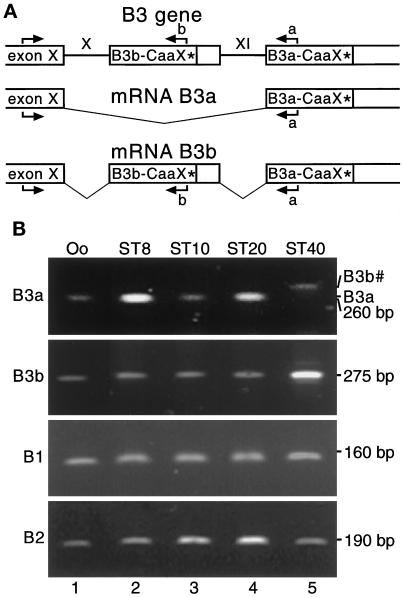
Figure 1.
Differential expression of two alternatively spliced lamin B3 mRNAs in Xenopus oocytes and embryos. (A) A scheme of the relevant region of the gene encoding Xenopus laevis lamin B3 and schemes of the two mRNAs that are generated by alternative splicing are shown. Exons are drawn as boxes, introns as lines, and spliced regions as thin V-shaped lines. Stars depict termination codons, and arrows mark the positions and the directions of the oligonucleotide primers used in RT-PCR. (B) Total RNA of oocytes (Oo) and different embryonic stages (ST8–ST40) was used for RT-PCR with primers specific for mRNA encoding lamin B3a (B3a), lamin B3b (B3b), lamin B1 (B1), or lamin B2 (B2). Ethidium bromide–stained agarose gels are shown. Size markers are indicated at the right. In panel B3a, the positions of amplicons resulting from amplification of either mRNA B3a (B3a) or mRNA B3b (B3b#) with B3a specific primer is indicated (see scheme in A for explanation).

