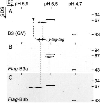
Figure 2.
Two variants of lamin B3 are expressed in Xenopus oocytes. Three nuclei each of noninjected oocytes (A) or oocytes injected with RNA encoding lamin Flag-B3a (B) or Flag-B3b (C) were manually isolated, and proteins were separated by IEF in the first dimension (IEF) and by SDS-PAGE in the second dimension (SDS). Lamins were detected after immunoblotting with the use of either mAb L6-5D5, which recognizes lamin B3 [B3 (GV)], or mAb M2, which recognizes the Flag epitope (Flag-B3a and Flag-B3b). The shift in isoelectric point of the two epitope-tagged lamins, which is due to the presence of the Flag epitope, is indicated by broken lines. Note the higher resolution of the IEF gel in the range between pH 5.7 and 5.8 caused by the nonlinearity of the pH gradient. Isoelectric points (averages) of standard proteins in the first dimension (left to right: phosphorylase b, BSA, and ovalbumin) are given at the top. The positions of the isoelectric variants of BSA and ovalbumin are indicated by brackets within each blot. The sizes of molecular mass markers in the second dimension are given in kilodaltons at the right.

