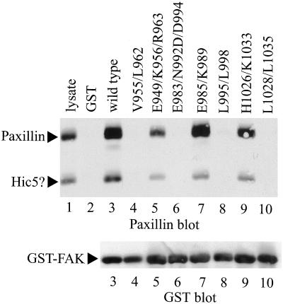
Figure 2.
Paxillin-binding activity of FAK mutants. Each mutant was expressed as a GST fusion protein and immobilized to glutathione agarose beads. CE cell lysate was precleared by incubation with GST alone immobilized to glutathione agarose beads. The cleared lysates were then incubated with GST (lane 2), GST-Hter (wild type) (lane 3), GST-FAKV955/L962 (lane 4), GST-FAKE949/K956/R963 (lane 5), GST-FAKE983/N992D/D994 (lane 6), GST-FAKE985/K989 (lane 7), GST-FAKL995/L998 (lane 8), GST-FAKH1026/K1033 (lane 9), or GST-FAKL1028/L1035 (lane 10). The beads were washed and bound paxillin was detected by Western blotting (top). Twenty-five micrograms of lysate was run as a control (lane 1). The paxillin band and a second reactive protein that is presumably hydrogen peroxide-inducible clone 5 are indicated by arrows. The blot was stripped and reprobed with a GST antibody as loading control for the GST-FAK fusion proteins (bottom).

