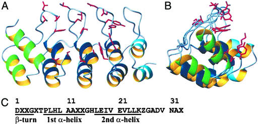
Figure 2.
Structure of the consensus AR protein E3_5. (A and B) Perpendicular views of E3_5 prepared with molmol (53). Ribbon representation of E3_5 showing the helices of the N-terminal, internal (consensus), and C-terminal repeats in green, dark blue, and light blue, respectively. The side chains of amino acids at randomized positions are highlighted in red. (C) The consensus AR sequence. X, any amino acid but C, G, or P; Z, any of the amino acids H, N, Y.

