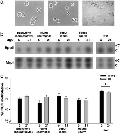
Figure 1.
Global genomic DNA determination by TLC. (a) Phase-contrast photographs of purified pachytene spermatocytes (Left), round spermatids (Center), and cauda sperm (Right) (×400). (b) Representative TLC plates of HpaII- and MspI-digested genomic DNA in germ cells and liver of young and old animals. The MspI-digested DNA reveals the relative content of cytosine (C) to m5C. (c) Graphical representation of quantified TLC results of germ cells and liver of young (black bars) and old (gray bars) animals. *, P < 0.05. Error bars represent ± SEM. Two-way ANOVA (n = 4–6 per age per group) revealed no significant effect of age, but methylation was significantly higher in liver when compared to pachytene spermatocytes (P = 0.019), round spermatids (P < 0.001), caput sperm (P = 0.005), and cauda sperm (P < 0.001).

