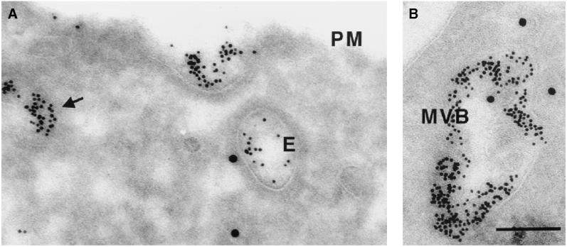
Figure 1.
Ultrastructural analysis of endobrevin localization. NRK cells were incubated for 15 min with BSA-gold (5 nm) before fixation and immunolabeling of endobrevin with the use of an affinity-purified rabbit serum and 15 nm protein A–gold (see MATERIALS AND METHODS). PM, plasma membrane; E, endosome of vacuolar type; MVB, multivesicular body. Note the association of endobrevin with tubulovesicular structures (A, triangle), with endosomal structures of vacuolar type (A, and with endosomes appearing as multivesicular bodies (B). These endobrevin-positive compartments are also positive for the endocytic tracer. Bar, 100 nm.

