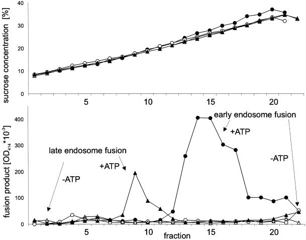
Figure 5.
Separation of early and late endosomes by continuous sucrose density gradient centrifugation after completion of the homotypic fusion reaction. Fusion reactions were carried out for early (circles) and late (triangles) endosomes in the presence (closed symbols) or absence (open symbols) of ATP and then loaded in parallel on top of 10–40% continuous sucrose gradients (see MATERIALS AND METHODS). At the end of the run, fractions of 0.5 ml were collected manually and analyzed by immunoprecipitation for the presence of fusion product. Each immunoprecipitate was assayed for HRP activity with the use of a photometric assay (bottom). The top panel shows the sucrose concentration of each gradient as determined by refractometry (data were obtained from a single experiment).

