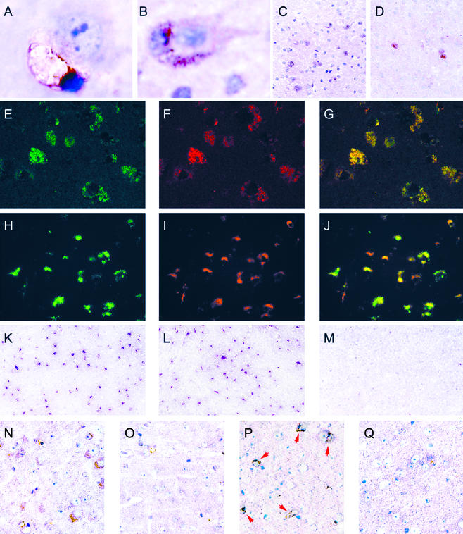
Figure 2.
Histochemical and immunohistochemical characterization of microglia in cortex. (A) G. simplicifolia isolectin IB4-positive microglia apposed to neuron; MPS IIIB, 7 months (×1,750). (B) MOMA-2-positive microglia apposed to neuron; MPS IIIB, 3 months (×1,150). (C and D) MOMA-2-positive cells seen at low power; MPS IIIB 3 months and MPS I, 14 months, respectively (×140). (E–G) Confocal images of sections stained with antibody against Lamp-1 (green), MOMA-2 (red), and merged; MPS IIIB, 6 months (×350). (H–J) Fluorescent images using antibody against ganglioside GM3 (green), MOMA-2 (red), and merged; MPS IIIB, 6 months (×260). (K–M) Images of sections stained with antibody against CD68/macrosialin, MPS IIIB, MPS I, and control, respectively, 3 months (×65). (N and O) Images of sections stained with antibody against IFN-γ; MPS IIIB, and control, respectively, 3 months (×250). (P and Q) Images of sections stained with antibody against IFN-γ receptor; MPS IIIB and control, respectively, 7 months (×250).

