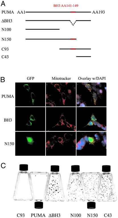
Figure 3.
Protein domains required for PUMA function. (A) GFP-PUMA expression constructs. GFP was fused to the N terminus of the indicated fragments of PUMA cDNA. (B) GFP-PUMA constructs were transfected into 911 cells and visualized through the fluorescence of GFP. MitoTracker red dye was used to visualize mitochondria. The N150 construct devoid of the C-terminal 43 residues had a diffuse staining pattern that was distinct from GFP-PUMA or GFP-PUMAΔBH3. (C) DLD-1 cells were transfected with the indicated constructs and selected with geneticin for 2 weeks before staining with crystal violet. There was no difference in colony formation after transfection with PUMA-ΔBH3 compared with transfection with the empty vector (data not shown).

