Figure 4.
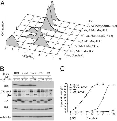
Biochemical changes during PUMA-induced apoptosis are Bax-dependent. (A) Cells with the indicated genotype were infected with Ad-PUMA or Ad-PUMAΔBH3 viruses, harvested at the indicated times, stained with CMXRos, and analyzed by flow cytometry to measure changes in mitochondrial membrane potential (Δψ). FL2 on the x axis reflects the intensity of the CMXRos signal. (B) Lysates from cells indicated genotypes infected with Ad-PUMA (wt) or Ad-PUMAΔBH3 (Δ) were analyzed by immunoblotting, using antibodies specific for the indicated proteins. The intact caspase 9 polypeptide is 46 kDa, whereas a degraded fragment of 37 kDa is detected in cells undergoing apoptosis. Tagged PUMA and PUMAΔBH3 proteins were detected by an antibody to HA. (C) DLD-1 cells were induced to express either PUMA or p53 in the presence or absence of the caspase inhibitor Z-VAD. At the indicated times, cells were assayed for apoptosis by fluorescence microscopy after staining with DAPI.

