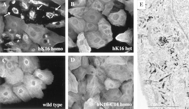
Figure 2.
Organization of keratin filaments in primary keratinocyte cultures. (A–D) Keratinocytes from wild-type, homozygous K16-C14 ectopic, and heterozygous and homozygous K16 ectopic keratinocytes were isolated and grown in culture for 72 h in standard calcium (0.2 mM) medium conditions and then immunostained to reveal keratin filament organization. (A) K16 homozygous cultures stained with the anti-K16 (1275) polyclonal antibody show striking alterations in filament organization in a subset of cells (arrows). No filament reorganization is observed in heterozygous K16 (B) cultures immunostained with anti-K16 anti-K16 (1275), or in wild-type (C) and homozygous K16-C14 (D) cultures immunostained for K17. Bar, 20 μm. (E) Similar homozygous K16 cultures were processed for electron microscopy studies. A subset of keratinocytes shows large electron-dense aggregates near the nucleus that are consistent with keratin aggregates (KA). Adjacent to these aggregates are short keratin filaments (KF). The mitochondria (M) and nuclei (N) in these cells are intact. Bar, 2 μm.

