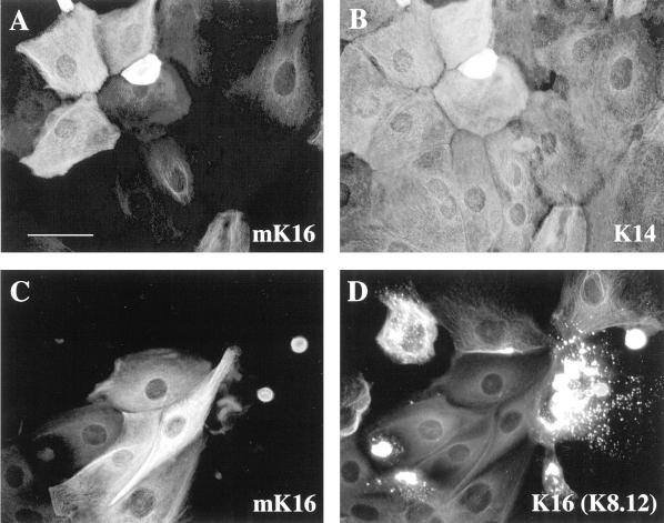
Figure 5.
Mutual exclusion of mK16 staining and keratin filament reorganization. Keratinocytes were grown for 72 h in 0.2 mM calcium, fixed, and immunostained. (A and B) Coimmunostaining of wild-type keratinocytes with an anti-mouse K16-specific antibody (A) and a monoclonal anti-K14 (LL001) antibody (B) shows that the mouse K16 antigen is expressed in a subset of keratinocytes in primary cultures. (C and D) Coimmunostaining of homozygous K16 keratinocytes with the anti-mouse K16-specific antibody (C) and monoclonal antibody K8.12 (D) as a marker of keratin aggregates shows that mouse K16 antigen cannot be detected in from K8.12-positive keratinocytes. Bar, 20 μm.

