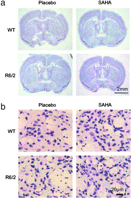
Figure 4.
Effect of SAHA on gross and cellular brain morphology. Frozen brain sections were cut from mice at 13 weeks of age, between Bregma 0 and 0.5 mm in the region of 0.26 mm and stained for Nissl substance with cresyl violet. (a) There is no marked change in gross morphology between R6/2 and WT mice in either treatment group. (b) Cellular atrophy is apparent in Nissl-stained sections from R6/2 brains. Treatment of R6/2 mice with SAHA resulted in Nissl staining more closely resembling that seen in WT mice.

