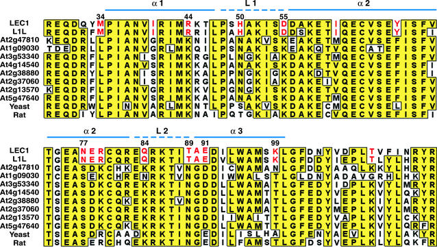
Figure 1.
Amino acid sequence alignment of the B domains of HAP3 subunits from Arabidopsis, yeast, and rat. Identical residues are shaded in yellow boxes. Residues specific to LEC1 and L1L are highlighted in red. The positions of α-helices and loops in the histone fold motif are indicated as continuous and dashed blue lines, respectively. Numbers indicate amino acid position within LEC1.

