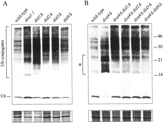
Figure 6.
Suppression by did mutations of the small ubiquitinated species that accumulate in doa4 cells and accumulation of ubiquitinated proteins in did single mutants. Extracts from a congenic set of strains were analyzed by anti-ubiquitin Western immunoblotting. Proteins were separated on a 16% Tricine gel; doa4 cell-specific species are marked with an asterisk. Monoubiquitinated species are detected poorly with the anti-ubiquitin mAb. The bottom panels show a section of the Coomassie blue–stained filters used for immunoblotting to indicate the relative loading of protein in each lane.

