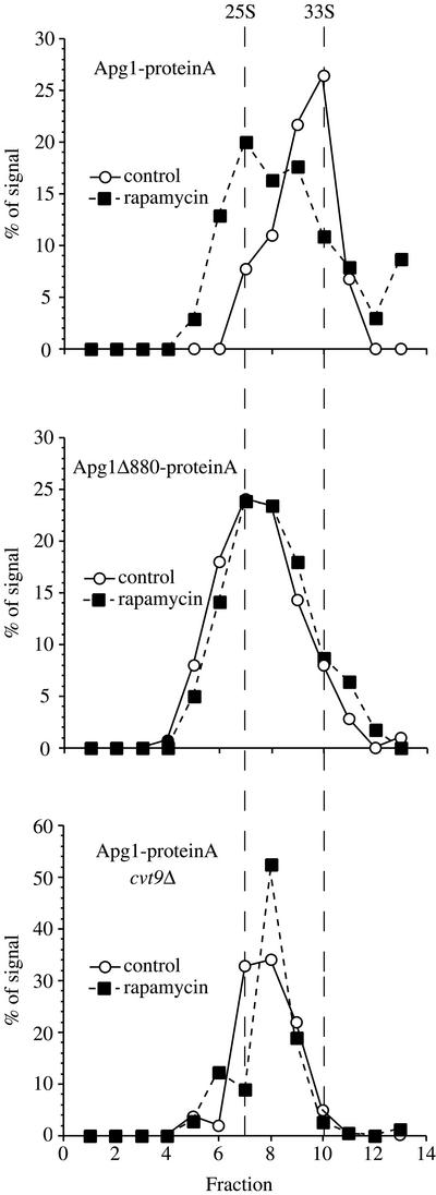
Figure 6.
Structural changes occur in Apg1 upon induction of autophagy and depend on Cvt9 and the C terminus of Apg1. The Apg1-protein A pep4Δ (HAY437), Apg1Δ880-protein A pep4Δ (HAY478), and Apg1-protein A cvt9Δ pep4Δ (HAY591) strains were grown to midlog in YPD and converted to spheroplasts. Spheroplasts were treated with or without rapamycin (0.2 μg/ml) for 15 min before lysis. Lysates were precleared by differential centrifugation at 100,000 × g for 15 min and loaded onto a 5–20% sucrose gradient. The gradients were centrifuged at 259,000 × g for 7 h, fractions were precipitated with 10% TCA and Apg1-prA was identified by Western blotting with anti-prA antiserum and quantified using a Bio-Rad Fluor-S MAX. The migration of the two peaks observed in wild-type cells is denoted by the vertical dashed lines (calculated to be 25S and 33S for the slow and fast moving components, respectively).

