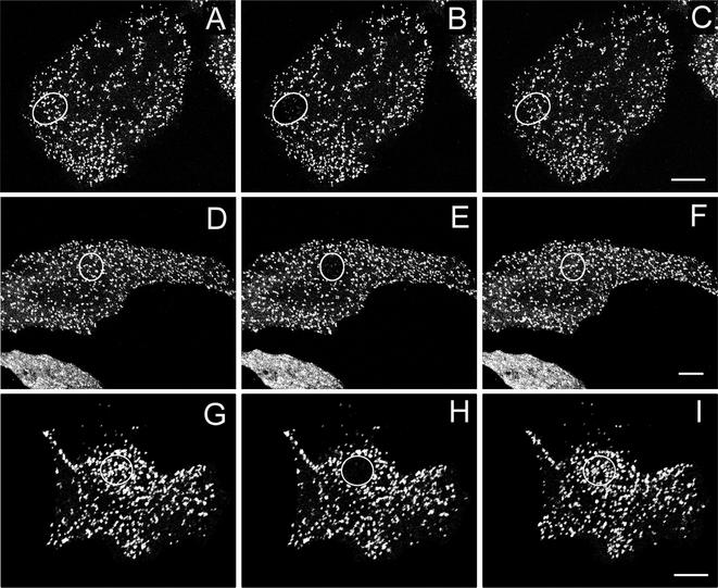
Figure 2.
Fluorescence recovery after photobleaching GFP-AP2 in HeLa cells at 37°C under different conditions. (A–C) Control HeLa cells. (D–F) HeLa depleted of cholesterol. (G–I) HeLa cells expressing K44A-dynamin. (A, D, and G) Images obtained directly before being photobleached. (B, E, and H) Images immediately after photobleaching. (C, F, and I) Images 2 min after photobleaching. The photobleached area is indicated in each figure. Bars, A–C, 6.5 μm; D–F; 9.5 μm; and G–I, 9 μm.

