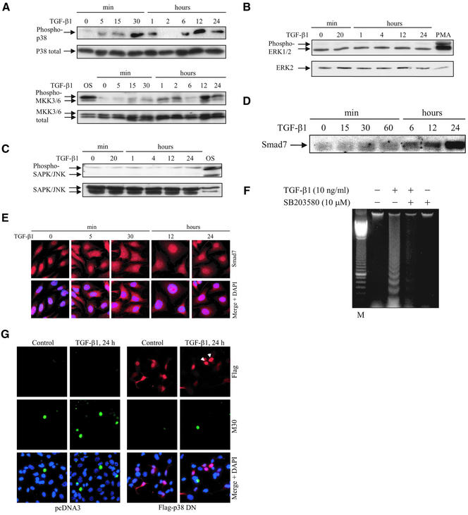
Figure 1.
Specific activation of p38 by TGF-β1 causes apoptosis. Time courses of phosphorylation of endogenous p38 and MKK3/6 (A), ERK1/2 (B), and SAPK/JNK (C), and expression levels of endogenous Smad7 (D) after TGF-β stimulation. Total cell lysates were prepared from TGF-β1–treated or untreated PC-3U cells and used for immunoblotting. In the top and bottom panels, the phosphorylated and the nonphosphorylated forms, respectively, of p38, MKK3/6, ERK1/2, and SAPK/JNK are indicated. Cell lysates from PC-3U cells exposed to 0.7 M NaCl for 30 min (OS) or 10 nM PMA for 20 min (PMA) were used as positive controls. (E) Subcellular localization of endogenous Smad7 in untreated PC-3U cells or PC-3U cells treated for 5 min, 30 min, 12 h, or 24 h with TGF-β, as investigated by immunostainings with antibodies against Smad7 and additional staining of the nuclei by DAPI. An overlay of both pictures (merge + DAPI) shows that Smad7 is predominantly localized in nuclei of untreated cells, whereas 5 min after TGF-β treatment an export to the cytoplasm is observed, followed by accumulation in the nucleus after 12 and 24 h. (F) Analysis of fragmentation of DNA isolated from PC-3U cells, TGF-β1–treated or not for 24 h in the presence or absence of SB203580. (G) Apoptosis of PC-3U cells, transiently transfected with pcDNA3 (control) and dominant negative Flag p38 (Flag-p38 DN), treated with TGF-β1 for 24 h, as analyzed by immunostainings with antibodies against Flag and the apoptotic marker M30. An overlay of both pictures with additional staining of nuclei with DAPI (merge + DAPI) showed a reduced number of M30-positive cells per field in cells expressing Flag-p38 DN after treatment with TGF-β1, compared with control cells. Note that cells expressing Flag-p38 DN, indicated by arrowheads, do not undergo apoptosis.

