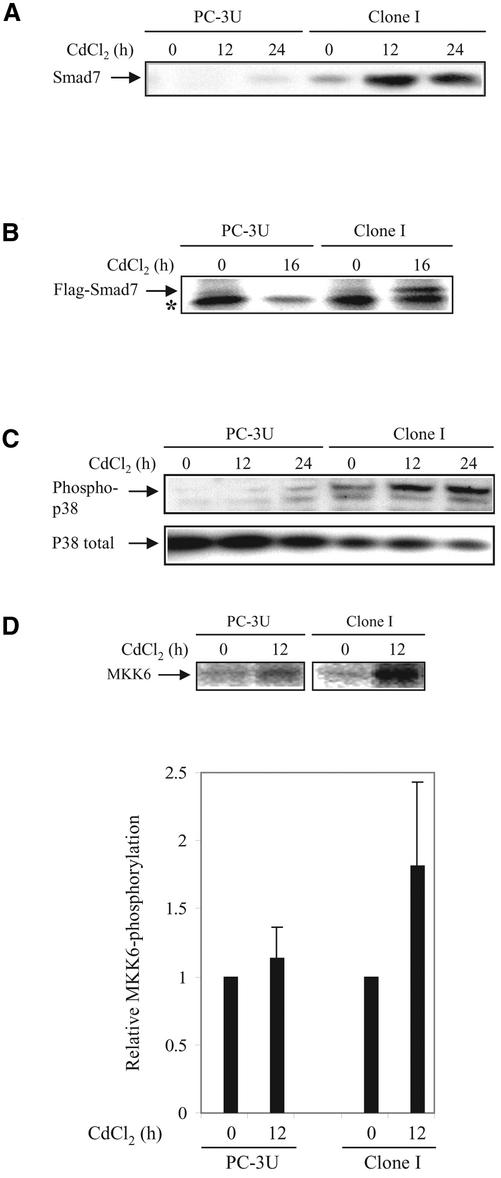
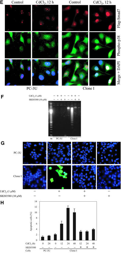
Figure 3.
Ectopic expression of Smad7 induces increased levels of p38 phosphorylation and apoptosis. (A) Time course of Smad7 levels in PC-3U and PC-3U cells stably transfected with pMEP-4 F-Smad7 (Clone I cells). Total cell lysates were prepared from wild-type PC-3U cells and Clone I cells, treated or not with CdCl2 and subjected to immunoblotting. (B) Ectopic expression of Flag-Smad7 is induced by CdCl2 stimulation (16 h) of Clone I cells as shown by metabolic labeling and immunoprecipitation with Flag antibody. A background band is indicated by an asterisk (*). (C) Time course of phosphorylation of endogenous p38. Total cell lysates were prepared from wild-type PC-3U cells and Clone I cells, treated or not with CdCl2 and subjected to immunoblotting. The phosphorylated p38 (phospho-p38) and the total p38 are shown in the top and bottom panels, respectively. (D) Activation of MKK6 by Smad7 overexpression is shown. PC-3U and Clone I cells were treated or not with CdCl2 for 12 h. Cell lysates were immunoprecipitated with anti-TAK1 antibodies and immunoprecipitates were subjected to in vitro kinase assay by using His-MKK6 as substrate. The phosphorylated proteins were resolved by SDS-PAGE and visualized by autoradiography (top). The mean relative values from three independently performed experiments including scanning electron microscopy are presented (bottom). (E) Immunofluorescence analyses of phosphorylated p38 in PC-3U cells and Clone I cells treated or not with CdCl2 for 12 h. Cells were also analyzed for Flag-Smad7 expression by immunofluorescence staining by using Flag antibodies. An overlay of both pictures with additional staining of nuclei by DAPI (merge + DAPI) reveals several nuclei with a colocalization of Smad7 and phospho-p38 in Clone I cells. (F) Analysis of fragmentation of DNA isolated from control PC-3U cells and Clone I cells treated or not for 24 h with CdCl2, in the presence or absence of SB203580. (G) Stainings with DAPI and TUNEL (fluorescein isothiocyanate) of control PC-3U cells and Clone I cells treated or not for 24 h with CdCl2 in the presence or absence of SB203580. Note the enhanced TUNEL staining of nuclei showing morphological criteria for apoptosis in cells overexpressing Flag-Smad7, which is inhibited by SB203580. (H) Apoptosis of PC-3U cells and Clone I cells treated or not with CdCl2, alone or together with SB203580, as revealed by immunostaining with the apoptotic marker M30; staining was quantified as described in MATERIALS AND METHODS. Note that the increase of M30 staining in Clone I cells treated with CdCl2 is reduced by SB203580, whereas only a minor apoptotic effect was detected in PC-3U cells after 24 h treatment with CdCl2.


