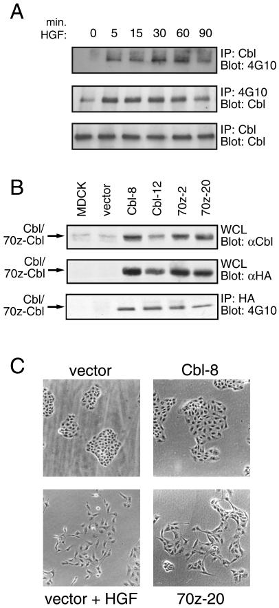
Figure 1.
Overexpression of 70z-Cbl in MDCK cells results in alterations in cell morphology. (A) MDCK cells were serum starved for 48 h and stimulated for the indicated times with 100 U/ml HGF. Equal amounts of cell lysate were immunoprecipitated with either anti-Cbl antibody (upper and lower panels) or anti-phosphotyrosine antibody (middle panel). The proteins were resolved by SDS-PAGE and immunoblotted with anti-phosphotyrosine (upper panel) or anti-Cbl (middle and bottom panels) antibodies. (B) Proteins from whole cell lysates from stable MDCK cell lines were separated by SDS-PAGE and immunoblotted with either anti-Cbl (upper panel) or anti-HA antibody (middle panel). HA-tagged Cbl proteins were immunoprecipitated with anti-HA antibody, separated by SDS-PAGE, and immunoblotted with anti-phosphotyrosine antibody (lower panel). (C) MDCK cell lines expressing vector, c-Cbl (clone 8), or 70z-Cbl (clone 20) were incubated for 24 h with or without 5 U/ml HGF, as indicated, and cell colonies were visualized by light microscopy.

