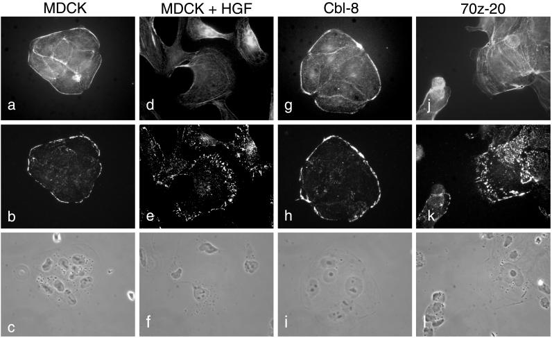
Figure 3.
Reorganization of actin and focal contacts in MDCK cells overexpressing 70z-Cbl. MDCK cells (a–c), MDCK cells stimulated with 5 U/ml HGF for 24 h (d–f), c-Cbl (clone 8; g–i), and 70z-Cbl (clone 20; j–l) were grown on glass coverslips for 24 h and treated with CSK buffer for 10 min. After fixation, the cells were double-labeled with phalloidin (a, d, g, and j) and anti-vinculin antibody (b, e, h, and k). Matching phase-contrast images are shown (c, f, i, and l). Photographs were taken at a magnification of ×63.

