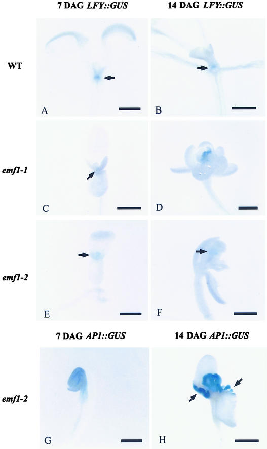
Figure 6.
Histochemical Localization of GUS Activity in 7- and 14-Day-Old Wild-Type and emf1 Seedlings Harboring the LFY::GUS or AP1:: GUS Transgene.
GUS activity is indicated by blue color. Bars = 0.5 mm.
(A) LFY::GUS wild type (WT) at 7 DAG, showing GUS activity at the shoot tip (arrow).
(B) LFY::GUS wild type at 14 DAG, showing GUS activity at the meristematic region of the shoot tip (arrow).
(C) LFY::GUS emf1-1 at 7 DAG, showing GUS activity at the shoot tip (arrow).
(D) LFY::GUS emf1-1 at 14 DAG, showing GUS activity at the shoot tip.
(E) LFY::GUS emf1-2 at 7 DAG, showing GUS activity at the shoot tip (arrow).
(F) LFY::GUS emf1-2 at 14 DAG, showing GUS activity in a patch of the carpelloid tissue (arrow).
(G) AP1::GUS emf1-2 at 7 DAG, showing GUS activity in the shoot tip, cotyledons, and hypocotyl.
(H) AP1::GUS emf1-2 at 14 DAG, showing GUS activity in the carpelloid structure and the papillae tissue developed at the base of the cotyledons (arrows).

