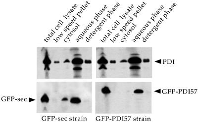
Figure 6.
Subcellular fractionation and partitioning of PDI and GFP chimeras. Cells expressing GFP-sec or GFP-PDI57 were mechanically broken in TKM buffer (150 mM KCl, 5 mM MgCl2). The total cell lysate was centrifuged at 1000 × g for 10 min to obtain the low-speed pellet. The supernatant was ultracentrifuged, yielding the cytosol fraction. The crude membrane pellet was subjected to Triton X-114 phase separation (aqueous phase and detergent phase). Equal amounts of each fraction, allowing direct comparison, were loaded on 12% SDS-PAGE under reducing conditions and analyzed by immunoblotting with the use of anti-PDI (upper panels) and anti-GFP (lower panels) antibodies.

