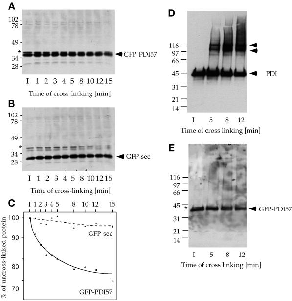
Figure 9.
In vivo cross-linking experiments in cells expressing GFP-sec or GFP-PDI57. Cross-links were performed with 106 intact cells with the use of 0.15% glutaraldehyde for 1–15 min as indicated (I, input). Samples were separated either on uniform 10% (A and B) or 4–15% gradient (D and E) SDS-PAGE under reducing conditions and immunoblotted with anti-GFP or anti-PDI antibodies. The 35-kDa GFP-PDI57 band decreased as a function of time (A), whereas GFP-sec was stable in these conditions (B). The band visible in A and B (asterisks) is a minor cross-reaction product of the anti-GFP antibodies. The resulting cross-link products were not resolved on the gels (see text). (C) Densitometric quantification of data in A and B. Samples from similar cross-linking experiments were separated on 4–15% gradient gels. Immunoblotting against PDI revealed the generation of two major complexes of ∼100 and 120 kDa (D), which did not contain detectable GFP-PDI57 (E). In addition, GFP-PDI57 was incorporated into heterogeneous cross-link products visible only as a weak smear (E).

