
Figure 3.
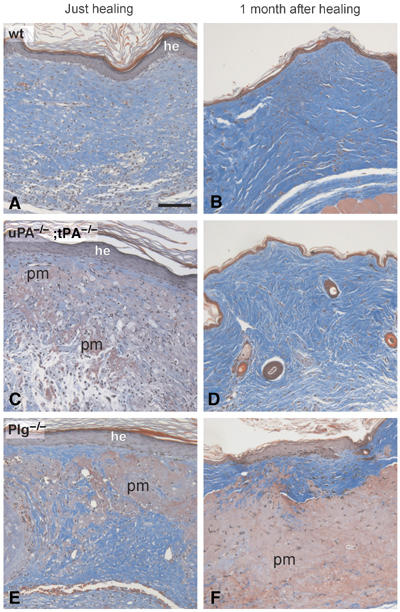
Analysis of skin wounds in wild-type, uPA;tPA double-deficient and Plg-deficient mice at the time of healing and 1 month post-healing Trichrome-stained sections of wounds from wild-type (A, B), uPA;tPA double-deficient (C, D) and Plg-deficient mice (E, F). Sections of tissue isolated just after healing reveal an intact, but hyperplastic epidermis (he), In wild-type mice (A), the provisional matrix has been completely replaced by granulation tissue, whereas in uPA;tPA double-deficient and Plg-deficient mice, larger areas of nondegraded provisional matrix (pm) were interspersed in the granulation tissue (C, E). At 1 month after healing, wild-type (B) and uPA;tPA double-deficient (D) mice all reveal a normal-looking epidermis and dermis, whereas in the Plg-deficient mice the epidermis is hyperplastic and the dermis is abnormal with abundant accumulations of undegraded provisional matrix (F). Bar: 60 μm.