
Figure 4.
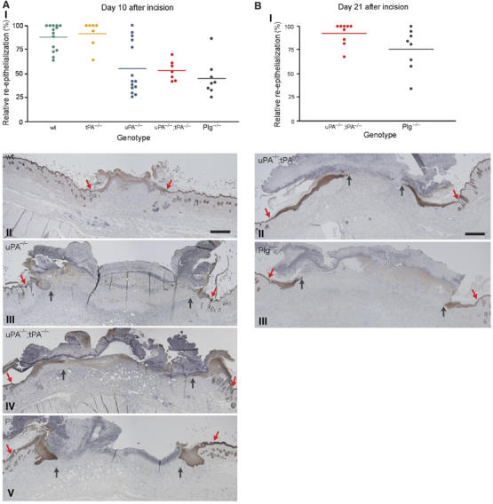
Morphometric analysis of wound re-epithelialization in wild-type, tPA-deficient, uPA-deficient, uPA;tPA double-deficient and Plg-deficient mice. (A) Scatter plot of the relative re-epithelialization of wounds day 10 after incision in wild-type mice, tPA-deficient mice, uPA-deficient mice, uPA;tPA double-deficient mice and Plg-deficient mice (AI). The relative re-epithelialization was determined as the distance from the wound edge to the front of the leading-edge keratinocytes, divided by the distance between the two wound edges. Examples of keratin-stained sections of day 10 wounds from wild-type mice (AII), uPA-deficient mice (AIII), uPA;tPA double-deficient mice (AIV) and Plg-deficient mice (AV). The red arrows mark the wound edge and black arrows point to the tip of the leading-edge keratinocytes. Bar: II–V ∼60 μm. (B) Scatter plot of the relative re-epithelialization of wounds day 21 after incision in uPA;tPA double-deficient mice and Plg-deficient mice (BI). Examples of keratin-stained sections of day 21 wounds from uPA;tPA double-deficient mice (BII) and Plg-deficient mice (BIII). Red arrows mark the wound edge and black arrows identify the tip of the leading-edge keratinocytes. Bar: II–III ∼60 μm.