
Figure 2.
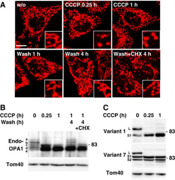
Processing of OPA1 after dissipation of membrane potential. (A) HeLa cells stained by MitoTracker were cultured in the presence of 20 μM CCCP for 0.25 or 1 h. After 1 h culture, CCCP was washed out and the cells were further cultured for 1 or 4 h with or without 1 mM cycloheximide (CHX). Images were obtained by confocal microscopy. Inset: magnified images. Scale bar: 10 μm. (B) HeLa cells were treated as in (A). Lysates obtained from these cells were subjected to immunoblotting using anti-OPA1 antibodies. (C) HeLa cells expressing OPA1-FLAG variants were cultured under the indicated conditions, and the cell lysates were subjected to immunoblotting using anti-FLAG antibodies.