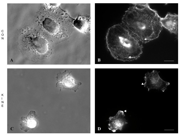
Figure 2.

Morphological analysis and actin organization of KINE and CON cells. Cells were grown in complete medium for 24 h before fixation and processing for phase contrast and epifluorescence microscopy. (A,B) CON cells form rosettes or clusters and appeared more spread and polarized compared to (C,D) KINE cells, which rarely cluster. Alexa 488-Phalloidin staining of actin shows more prominent stress fibres in (B) CON (arrows) compared (D) KINE cells. KINE cells, however, possess actin-rich membrane ruffles at the leading edge (arrowheads) absent in CON cells. (Bars = 5 microns).
