
Figure 2.
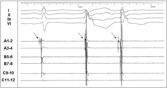
Intracardiac electrograms during spontaneous ventricular ectopy in a patient with Andersen-Tawil syndrome. Displayed are surface leads I, II, III, V1, and intracardiac electrograms recorded from the anterior left ventricle. The first sinus beat is followed by 2 successive ventricular ectopic beats, each with a different morphology. The arrows indicate Purkinje potentials preceding the local ventricular electrograms in sinus rhythm and the ectopic beats. The first ectopic beat is from an adjacent Purkinje site - hence the late Purkinje relative to the QRS. (Figure courtesy of Dr. Prashanthan Sanders, Dr. Frederic Sacher, and Dr. Michel Haissaguerre, Hôpital Cardiologique du Haut-Lévêque, Bordeaux, France)