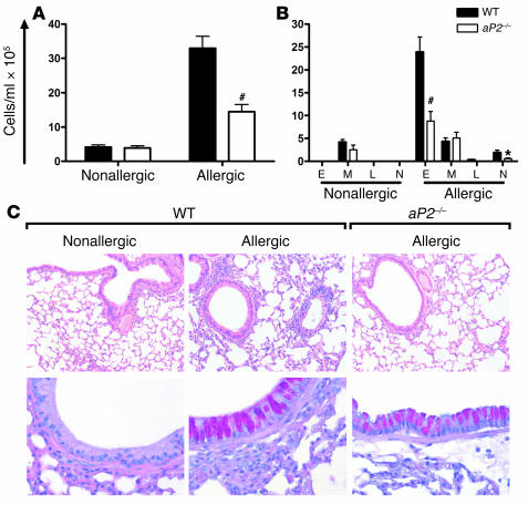
Figure 5. aP2 deficiency ameliorates allergic lung inflammation and airway eosinophilia.
WT or aP2–/– mice were sensitized and challenged with OVA for airway inflammation (Allergic) or with PBS as a control (Nonallergic). Total (A) and differential (B) cell counts from BAL. E, eosinophil; M, monocyte/macrophage; L, lymphocyte; N, neutrophil. Data represent mean values ± SEM for n = 10 nonallergic and n = 12–15 allergic WT or aP2–/– mice from 2 experiments. *P < 0.05, #P < 0.0005 compared with WT. (C) Histological examination of lungs stained with H&E (upper panel; original magnification, ×100) or Alcian blue–PAS (lower panel; original magnification, ×400) for mucus. Magenta staining is indicative of mucus.

