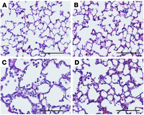
Figure 2. Histopathology of the lungs of mice treated with CVT-6883.
Lungs were collected from postnatal day 38 mice and prepared routinely for sectioning and H&E staining. (A) Lung from an ADA+ mouse treated with vehicle. (B) Lung from an ADA+ mouse treated with CVT-6883. (C) Lung from an ADA–/– mouse treated with vehicle. (D) Lung from an ADA–/– mouse treated with CVT-6883. Sections are representative of 6–8 different mice from each treatment group. Scale bars: 100 μm.

