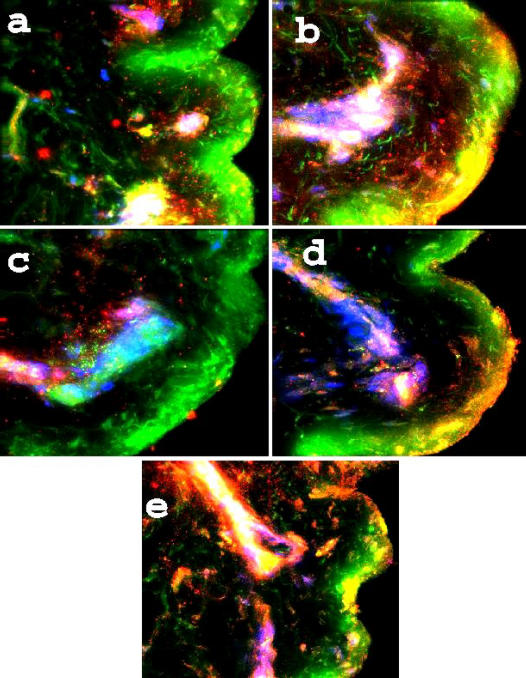
Figure 2.
Antimicrobials in burned skin. A 5-panel collection of fluorescence images in burned skin samples. Magnification is 400×, and the probes are as used in Figure 1. Of note is the HBD-1 associated with dermal structures, not only in the upper levels but also with eccrine epithelia (a), HBD-2 associated with gland epithelia (b), HBD-3 in the lower papillary dermis (c), HNP along a hair follicle and on the upper, denatured keratin layer (d), and LL-37 very apparent via the bright yellow colocalization (green + red), along a sweat duct and deposited on the corneum at the point of exit.

