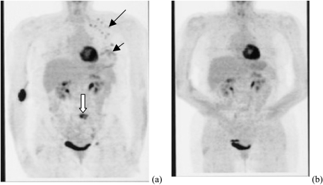
Figure 3.
PET images of a patient who presented with a diffusely swollen breast. Core biopsy revealed a high-grade infiltrating ductal cancer. (a) Image acquired before chemotherapy. The small black arrow indicates the diffuse involvement of the breast. The longer black arrow indicates positive lymph nodes (black dots). The white arrow displays an area of metastatic disease (epidural). (b) PET image acquired after eight cycles of chemotherapy, before definitive surgery. No evidence of disease remains.

