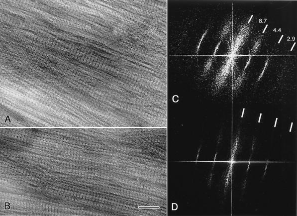
FIG. 2.
Transmission electron micrographs of negatively stained bundles of purified cytoskeletal ribbons that are densely packed and relatively well aligned; area A is thicker and less ordered than area B. Computed diffractograms of the ribbon bundles shown in A and B are presented in C and D, respectively; sharp meridional reflections corresponding to the axial repeat along the fibrils are evident. Lateral order is less preserved in the bundles, as indicated by the diffuse reflections. The axial repeat is ∼8.7 nm in both diffraction patterns. Bar, 50 nm.

