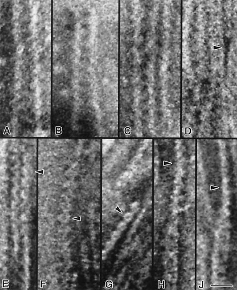
FIG. 5.
Transmission electron micrographs of negatively stained single and paired fibrils. Single fibrils ∼5 nm in width are shown in A to D. As two fibrils approach each other (panels A to D), the gap between them becomes filled with pools of stain (e.g., panel D, arrowhead) separated axially by ∼8 nm; two closely associated fibrils are seen in panel D (top). In this panel, two fibrils are seen to dissociate towards the lower part of the figure. This pattern of staining may indicate the presence of a periodic, concave structural element along the fibril that is prone to accumulate stain. The pairing of ∼4- to 5-nm fibrils into wider ∼9- to 10-nm structures is illustrated in panels E to G; pairs of serrated fibrils are visible in panels E and G. Chains of ring-like structures ∼9 nm in width and separated by ∼8 nm in the axial direction are shown in panels E to G (arrowheads). Twisted pairs of fibrils are illustrated in panels H and J; rings ∼10 nm in width are visible in the lower parts of these panels. At the center of both panels, pairs of fibrils are seen to twist, and at the top they are observed in side view (arrowheads) with the same axial repeat but with a width of only ∼5 nm. Bar, 20 nm.

