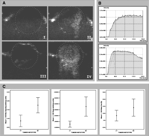
Figure 5.
In vivo assessment of BB1 and A17 tumor vasculature by CEUS with SonoVue contrast agent. CEUS showed that A17 tumors had a lower level of vascularization than BB1 tumors. Signal enhancement after contrast agent injection was less evident in A17 (A, I: precontrast; II: postcontrast) than in BB1 (A, III: precontrast; IV: postcontrast) tumors. Two representative time-intensity curves show differences between time course contrast enhancement observed in A17 (B, top panel) and BB1 (B, bottom panel) tumors. BB1 tumors had a reduced maximal intensity peak (Mi), delayed slope (S), and reduced wash-out (Wo) in comparison with BB1 tumors (C; see also Table 1).

