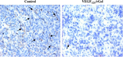
Figure 6.
Detection of Flk-1/VEGFR-2 on the vasculature of metastatic lesions by the anti-VEGFR-2 antibody, RAFL-1. Frozen sections of lungs from mice treated with VEGF121/rGel or free gelonin stained with monoclonal rat antimouse VEGFR-2 antibody RAFL-1 (10 µg/ml). RAFL-1 antibody was detected by goat antirat IgG HRP, as described under the Materials and Methods section. Sections were developed with DAB and counterstained with hematoxylin. Representative images of lung metastases of comparable size (700–800 µm in the largest diameter) from each treatment group are shown. Images were taken with an objective of x20. Note that the pulmonary metastases from the VEGF121/rGel-treated group show both reduced vessel density and decreased intensity of anti-VEGFR-2 staining compared to control lesions.

